We have newly re-engineered our original 3D viewer for use in medicine and dentistry, and have completely redesigned the user interface.

Our 3D viewer system for CT and MR images, which we have been developing since 2016 for use in medicine and dentistry, has now been redesigned with a completely new user interface.

In addition, several new features and improvements have been implemented this time. We will continue enhancing the functionality of this system in the future.

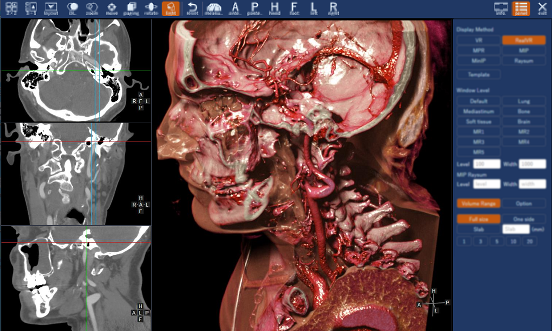
The top toolbar of the 3D Viewer is designed with buttons that are easy to understand at a glance. The right side panel for manipulating MPR and volume rendering images has been redesigned to be sleeker and easier to operate.

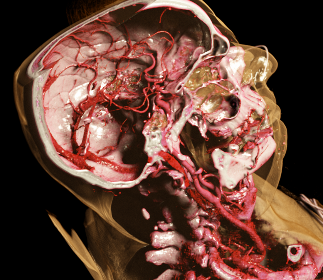
This high-impact image here is a volume-rendered image from our new 3D viewer with our photorealistic 3D rendering engine.

This image realistically depicts the anatomical structures of the arteries and each bone in the head and neck region. The image also clearly shows how the internal carotid artery connects from the bottom to the top through the skull base to arteries in the brain.

Here is a movie of the operation screen of our viewer displaying a dental cone-beam CT scan data.
Operations such as changing colors, adjusting the position of virtual light sources, rotating shapes, and zooming in on shapes can be performed quickly and easily.

The 3D viewer screen shown above is an example of a 2 x 2 layout and template panel (right side of the screen).

When in normal volume rendering mode or photoreal volume rendering mode, the 3D image can be quickly switched and displayed according to the part of the body, etc., by selecting the icon button on the template panel.

The images above are cases of a chest CT and a dental cone-beam CT.

The image on the left shows a three-dimensional cross-section of the right lung at the location of the mass, which clearly shows the state of spread of the mass within the lung area. The middle image shows the appearance of the lungs in the same case. The pleura can be seen to be pulled by the mass.

The two images on the right are volume-rendered images from cone-beam CT scan. In the image below, the left intra mandible is shown in transparency, allowing us to grasp the dental structure and the position of the tooth root in relation to the mandibular canal. Interactive, individual tooth-focused viewing and observation can be performed in real time. It can also measure the distance between the root and mandibular canal on the cross section.

News  Posted:2023.09.07  Last Updated:2024.06.06